I have seen papers using GFAP staining to assess satellite glial cell activation (gliosis) - my question is: is GFAP only expressed during SGC activation, or is it expressed at basal levels and then there is an increase in GFAP abundance during SGC activation?
I ask because I see several papers showing essentially no GFAP staining in naïve conditions in the DRG.
With many experimenters presenting the data as % of neurons surrounded by GFAP-immunoreactivity. The number of neurons surrounded by GFAP signal is usually <20% for controls, and then significantly increases after injury.

Nice images! I think you’re finding biological reality. @tberta will know the answer to this. I know in the spinal cord people say astrocytes are GFAP+ when reactive, but I think we all know there is lots of GFAP+ cells even in naive mice.
We have tested several GFAP Abs and none really worked well, and we always go back to the old but still the best MAB360 ( Anti-Glial Fibrillary Acidic Protein Antibody, clone GA5 ascites fluid, clone GA5, Chemicon® | Sigma-Aldrich (sigmaaldrich.com)).
Using this Ab, we have showed that naïve SGCs do not express GFAP (or only a few) and this is in agreement with our and other single-cell data.
See an example of the IHC using the MAB360 ( 1/500, BSA1%, vehicle ← vs nitroglycerin → ).

This is the antibody I am using - https://neuromab.ucdavis.edu/datasheet/N206A_8.pdf
I worked with this ab a lot when I worked for this company. Its KO validated via western and IHC, its very sensitive, works super great.
I am just wondering if this is an epitope difference? Or why does this staining look so robust in DRG in naive animals? I will order your recommended ab and compare. This is in mouse spinal cord:

Antibodies - what a puzzle?!# I don’t have the answers to your questions, but I am pretty sure that GFAP is very low expressed if not at all in naïve SGCs.
Here is some single cell data from our recent preprint

SGC also express glial fibrillary acidic protein (GFAP) and similarly to astrocytes, GFAP expression is increased under pathological conditions, which can have a protective function (Woodham et al, 1989). In mouse, Gfap is one of the top upregulated genes in SGC upon nerve injury (Avraham et al, 2020)(Christie et al., 2015; Fenzi, Benedetti, Moretto, & Rizzuto, 2001). However, in uninjured DRG, the distribution of Gfap was relatively low, with ∼20% expression in rat, 1.5% in mouse and undetectable levels in human DRG (Fig. 2d).
So the question is, which antibody is telling the truth? Probably the MAB360. But I agree @sshiers your antibody definitely looks like it’s binding SGCs and astrocytes. But is thsat GFAP or some other target? I feel like some RNAscope would be helpful here. @tberta have you done GFAP RNascope on mouse?
I did not, but it may be helpful. One last note, Gfap expression is developmentally regulated as GFAP-cre mice crossed with tdTomato-loxp mice show a robust signal in almost all SGCs!
I just don’t know how it could be seeing a different target. Its a monoclonal ab which only sees a specific epiptope which is usually ~20 amino acids and is knockout validated. Science is confusing sometimes  . I also used an isotype specific secondary to preference the IgG1 subtype since H&L secondaries can be IgG2b biased.
I am going to give MAB360 a shot. Its going a little out of the scope of the project goals but I am super curious.
Oh interesting - what is the cut off for the age when GFAP expression dies down? Maybe my mice are too young
Unfortunately, I don’t know when the GFAP expression peaks or weans off. Most of our IHCs are done in 6-12 weeks old mice. I think that increase in GFAP is a good indicator for reactive SGCs, but if you are interested to just label these cells GS and FABP7 are excellent cell markers for mouse and human SGCs.
I see you got many responses already. From my experience with gut, a sub-population of glia will always express decent amount of GFAP that can be increased when reactive. Same applies to S100b. I’m not sure about how it looks like in other peripheral tissue, though I’ve seen others showing nice GFAP under physiological conditions too.


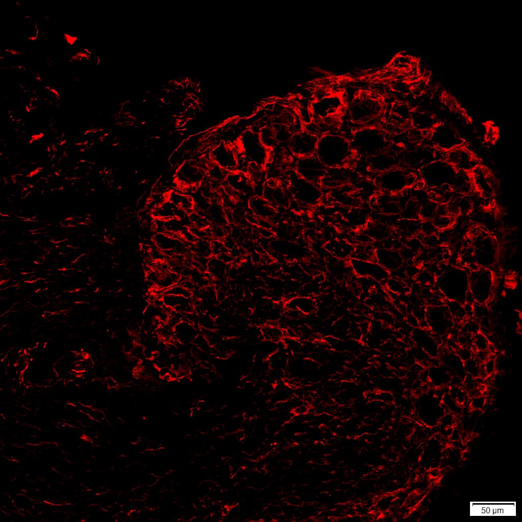
Thanks for sharing… Hmmm. MAB360 has less perimeter staining around neurons (SGCs?) but a lot of signal in that axon bundle. What’s up with that? Weird.
N206A/8 looks like it’s in SGCs, but even in naive SGCs… Which they say isn’t supposed to happen.
From our last discussion, some commentary here
Thanks for coming back and sharing. If you want just an SGC marker, use Fabp7.
It’s excellent.
Here it is in human DRG, 1:500. It’s beautiful.

If you’re looking for ‘reactive’ astrocytes, then I guess GFAP is what you can use. Try the MAB360
I think we may have gotten a bad lot of that FABP7 ab - it doesnt look good for us. ![]()
We were trying to label activated SGCs but a pan SGC marker is also useful.
Hmm. Yeah perhaps. That antibody has been very reliable, and I’m using an aliquot that has been abused (multiple freeze thaws) ![]()
We use PFA fixed tissue (4%, 24 hours). Shouldn’t make too much of a difference, but maybe? Sometimes I try antibodies you’ve used beautifully and don’t get the same staining, for example, CGRP. For some, these fixation/pre-treat conditions likely make a difference, as we’ve commiserated about before.